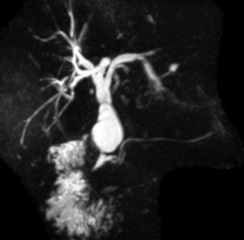
Choledochal Cyst
Choledochal cyst is a congenital condition wherein the bile duct abnormally dilates to form cystic structures. This can happen in the ducts outside the liver, the ducts inside the liver, or both.
Choledochal cyst is a premalignant condition. Approximately 60% of this condition is identified in the first decade of life, whereas 20% of it is identified during adulthood.
Choledochal cysts can be classified into five main types according to Todani classification:
- Type I is the most common choledochal cyst, which develops in the common bile duct (CBD) outside the liver.
- Type II develops as an isolated protrusion from the common bile duct; it is also known as bile duct diverticulum.
- Type III, or choledochocele, develops in the region where the bile duct meets the pancreatic ducts.
- Type IV develops as multiple cysts/dilations that occur in the intra- or extra-hepatic bile duct branches.
- Lastly, type V, also known as Caroli’s disease, develops as cystic dilations in the intra-hepatic biliary ducts.
Symptoms:
Choledochal cysts often exhibit the following symptoms:
- Jaundice
- Upper abdominal pain
- Cholangitis
- Pancreatitis
- it is often identified incidentally from imaging performed for unrelated conditions.
Treatment:
CT and MRI scans are commonly used to correctly diagnose a choledochal cyst. The choledochal cyst patient who presents with obstructive jaundice or cholangitis will require an Endoscopic retrograde cholangiopancreatography (ERCP) with stenting to drain the obstruction before surgical resection.
The surgical treatment varies according to the different Todani classification types e.g.
Type I: the bile duct can be surgically removed and reconstructed to aid the proper functioning of the bile flow.
Type III: can be treated with ERCP with sphincterotomy to open up the cyst.
Type IV: The bile duct has to be excised together with liver resection to remove all involved areas.


